Sunday

Bones

Do you know how many bones does a baby have? It is a blind belief that babies have fewer bones than adults. But here the facts about bones are totally different. In fact, the children have more bones than adults, summing up to 350! Mostly, they are in the form of cartilage and might not be stronger. Whereas adults have only 206 bones in their body. It is one of the amazing facts about bones that more than hundred bones disappear as a baby grows to become an adult.

The bones of babies are softer and flexible which need careful handling. When the cartilage and bones merge together, it constructs a stronger bone. In case of fractures and damages the concerned person needs to go to the doctor. Meanwhile, there are possibilities to set their bones immediately. The skeletal structure is so important for a man and there should be proper practice of body movements.

Do you know how many bones does a baby have? It is a blind belief that babies have fewer bones than adults. But here the facts about bones are totally different. In fact, the children have more bones than adults, summing up to 350! Mostly, they are in the form of cartilage and might not be stronger. Whereas adults have only 206 bones in their body. It is one of the amazing facts about bones that more than hundred bones disappear as a baby grows to become an adult.

The bones of babies are softer and flexible which need careful handling. When the cartilage and bones merge together, it constructs a stronger bone. In case of fractures and damages the concerned person needs to go to the doctor. Meanwhile, there are possibilities to set their bones immediately. The skeletal structure is so important for a man and there should be proper practice of body movements.

The spine bone acts as the base and anchor for all other bones, which is vital for the skeletal framework of the body. There are 33 spine bones which are joined and work together to form the vertebral column. The 24 bones, to the top, are moveable with the remaining nine bones are fused together. The less flexible joints in the spine bones provide limited freedom of movement.

The bones in general can be classified into two types namely the compact bones and the trabecular bones. The compact bones are the denser bones, which cover 80 percent of the total body bone mass and the trabecular bones are spongy in nature. They cover the remaining 20 percent.

⇛ The largest bone of the human body is the femur which you can find in the thigh. And the smallest bone is the stirrup which forms the ears.

⇛ Everybody should wear a helmet and protective pads or masks, while driving or involving in any sports activity.

⇛ To support the bones, consume the daily food items with balanced percentage of calcium and vitamin D content.

⇛ By practicing the weight bearing exercise like walking, jogging, climbing, dancing, and running there will be expansion and regular growth to strengthen your bones.

⇛ You should avoid alcohol consumption and smoking which results in the bone diseases like osteoporosis.

Adrenal Gland

The glands responsible for the production of adrenaline are known as adrenal glands. The hormones help you cope with situations of extreme stress, danger or excitement. They not only produce adrenaline, but several other hormones as well. These hormones are important for different functions of our body.

Adrenaline and noradrenalin are notable hormones of adrenal glands. Another main class of hormones is that of corticosteroid hormones. To know completely What is Adrenal Gland, you also need to know about the structure and location of these organs of the body as well.
 Concerning adrenal gland location, a pair of them appears at the top of the kidneys. Therefore, you can also call them the suprarenal glands. Here, the one to the right looks like a triangle. On the other hand, the left gland exists in a semi-lunar shape. However, each of them consists of two main parts. The outermost part is the cortex. It is responsible for the secretion of corticosteroid hormones. Similarly, the inner part is the medulla. It produces adrenaline and noradrenaline.

Adrenal glands function is to produce corticosteroids. The pituitary gland stimulates them for work. In turn, the pituitary is stimulated by the hypothalamus. In other words, the secretion of hormones is controlled by the pituitary gland and the hypothalamus.
Cancer of the adrenal glands can lead to over-production of aldosterone. Specifically, you may term such a condition as hyperaldosteronism. An individual experiences fatigue. Meanwhile, they suffer from several other adrenal gland symptoms. They include headache, high blood pressure and weakness of the muscles. In addition, paralysis may occur in extreme cases.

To sum up, it is easy to prevent the disorders of the suprarenal glands. Firstly, you should try to avoid the excessive use of steroid drugs. Moreover, go for a frequent examination of the adrenal glands. For example, scans can help to detect any development of cancer and tumors at an early stage. The information about ‘what is adrenal gland’ will be very useful for you.

Thursday

Hypothalamus

As you can see in the hypothalamus pictures, it measures about the size of an almond or pearl. It is an extremely important part of brain in human beings and other higher animals. Hypothalamus constitutes one of the four major components of diencephalon, while the other three are: thalamus, epithalamus and subthalamus. It is intimately associated with the function of autonomic & endocrine systems, formation of behavioral patterns, temperature control, reproduction, and so on.

With the help of hypophysis
, the hypothalamus develops a connection between endocrine system and nervous system in the body. A number of medical conditions have been found associated with this part of the interbrain that may arise out of various factors, and include nutritious, infectious, neoplastic and inflammatory disorders that are manifested by severe symptoms in the victim. As these problem areas are located inside the human brain, the diagnosis and treatment is not easy. However, if identified at an early stage, the symptoms can be relieved through medication.

The hypothalamic region in the posterior part of forebrain (diencephalon) initiates, facilitates and accomplishes a number of vital functions in the human body including hunger, circadian cycles, fatigue, sleep, thirst, attachment behaviors and parenting. The neurohormones (chemical substances of the nervous system) secreted by the organ are transferred to the pituitary gland that either inhibit or stimulate its secretary activities. Some of the vital functions of hypothalamus can be summarized.

It controls the secretion of hormones from the hypophysis which is an important part of the brain and master secretory organ in your body. Pituitary is associated with the release of eight essential hormones in the body; two of which are synthesized by the hypothalamus, while the remaining six are produced locally. Some of the other endocrine and exocrine functions of hypothalamus include the control of autonomic nervous system, maintenance of homeostasis, balance of emotions, regulation of hunger & thirst and check over the thermostat of the body.
Malnutrition, inflammation, neoplasm, infection or any other condition of this structure not only adversely affects its functions but also causes severe symptoms in the victim. Such signs need to be addressed as soon as possible. Originating from the adjacent structures, like optic chiasm, hypophysis or optic nerves, the benign or malignant tumors spread to affect the organ and lead to hypothalamic neoplasms. The clinical manifestations of the disorder include loss of vision, precocious puberty, macrocephaly and developmental delay. The frequently recommended treatment measures of the tumors include radiations and surgical therapy.

Hypopituitarianism is another ailment of brain in which pituitary gland and hypothalamus are integrated with each other, thus inhibiting the hormonal secretions of hypophysis and disturbing all the vital functions of the body that are regulated by the chemical substances produced therein. In this case, your health care provider may suggest hormonal replacement therapy as the disease results in the hormonal insufficiency in the body.

Brain


What a miraculous thing human brain is! It is one of the most powerful and marvelous creations of the Nature. It is known to have countless mysteries, the majority of which are still undiscovered and least understood. Your brain is a highly complex network of billions of neurons that render it the ability to think, feel, process information and produce responses. The question “What makes brain to initiate and accomplish the process of thinking” has not yet been answered properly. There are also a number of other related questions to which the researchers are trying to provide convincing answers. It has recently been discovered that, contrary to the earlier assumption, brain is not hardwired and can be changed or trained according to one’s will! Are you aware of the mind-blowing facts about your master organ? Well, it is high time to make yourself acquainted with some amazing realities about the human brain. This platform is specifically devoted to provide the readers with information that will not only be useful but also engaging and entertaining. Meanwhile, we also aim to keep you updated with the latest discoveries pertaining to the health, working and enhancement of brain.

Tuesday

Lymph Nodes


They behave like mechanical filters. The job of these mechanical filters is to kill and eliminate microbes present in the blood stream. The white blood cells (WBCs) or lymphocytes are produced by the lymph nodes. On the other hand, WBCs kill bacteria and viruses entering the body.

Lymph nodes are very small organs, measuring only about 1-2 cm in size. They form a network throughout the body. There are about 500 lymph nodes spread across various organs of the human body.

A lymph node gets its particular name from its location and relation with the body organs. For example, the cervical lymph nodes are present in the neck or cervical region of the body. They protect the tonsils and the pharynx. Here you should note that swollen cervical lymph nodes is an indication of the lymphatic system disease.

Lymph nodes in armpit are the axillary lymph nodes. Their job is to drain the chest region. You will find the mediastinal nodes near the sternum and the lung sacs. Meanwhile, for the drainage of the abdominal organs, there are the mesentery nodes.

Lymphatic System a Sewerage System: It won’t be wrong to compare the lymphatic system to a sewerage system. It has a networks of pipes to drain waste from every single body cell. The pipes in this system are defined as lymph vessels. The job of these vessels is to transport clear interstitial body fluid, that is, the lymph.

The Function of Lymph: The lymph is a watery fluid that flows in the lymphatic vessels. Its function is to help in effective draining mechanism.

Coordination of Drainage Mechanism: It is the muscles movements that coordinate the entire mechanism of draining. It is particularly because the lymphatic system has no specific pumping organs.

Bulging of Lymph Nodes: The lymph nodes bulge out in the presence of an infection. It is because, in case of an infection, they have to work hard to get rid of it.

Complementary Organs of Lymph Nodes: There are many organs in your body which protect the lymph nodes. They include spleen, tonsils, thymus and the adenoids. In addition to playing a protective role, these orangs are also complementary to the lymph nodes to form the immune system of body.

Lymphatic Circulation: About 2 liters of lymph fluid circulate in the body every day. During circulation, the lymph collects, destroys and eliminates foreign agents. Meantime, it also deals with the disease causing microbes.

Types of Lymphocytes: There are two types of lymphocytes or white blood cells that the lymph nodes produce. These are the B & T lymphocytes. The B-lymphocytes target bacteria and some viruses. Whereas the T-cells attack viruses in your body.

Monday

Parathyroid Gland

A number of vital functions in human body are controlled by biochemical substances, called hormones, which are chemical messengers released by either ductless or ducted glands directly into the blood stream or through ducts. Of all the hormone secreting glands in the body, the parathyroid gland is the smallest which is embedded in the thyroid gland or located on the rear side of it in the neck. There are four parathyroid glands located on the posterior side of the thyroid, measuring only about the size of a rice grain.
Because of the very small size, these endocrine glands weigh as low as 20mg or as high as 40mg. Usually, these are four in number, arranged in two pairs-one above and the other below-but some individuals have also been reported to have 6, 8 or even more. The two glands lying towards the head are termed as the superior parathyroid glands, while the other two lying on the lower side are known as the inferior parathyroid glands. Though the parathyroid glands are named after their close proximity with the thyroid gland, their functions are completely different from that of the neighbouring endocrine organ. Nervous and muscular systems are very sensitive to the concentration of calcium in the body that has to be maintained in a narrow range for their proper functioning. The hormone secreted by parathyroid gland (named after the gland as 'parathyroid hormone' or simply "parathormone") is responsible for the maintenance of phosphate and calcium homeostasis in the body, and is functionally opposed to calcitonin.

Though there are no pills or medications to cure the diseases of parathyroid gland, surgery and some other treatment measures have shown promising results.

Most of the parathyroid problems present themselves in their advanced stages, and some patients will not complain of any symptoms, until they are identified during routine diagnostic procedures.

Often the medical fraternity get confused with the diseases of the parathyroid gland and the blood calcium levels, as neither of these the conditions (diseases and calcium levels) have ever been found to synchronize with each other.

Most of the patients suffering from parathyroid disease have fluctuating blood calcium levels both in blood and bony structures.

The main function of the Parathyroid gland is to control the blood calcium levels within the range of 9-10 mg/dl which is done by the gastrointestinal absorption of calcium and its conservation in kidneys. It stimulates absorption of calcium from food and restricts kidneys from excreting excessive calcium in urine. If the blood calcium levels are low, the hormone would erode the bones to manage optimal blood calcium levels, thus making the bones weak and easily breakable. The gland also checks and regulates the levels of calcium deposition on the bones and their density.
Calcium not just makes the density of the bones and frames the skeletal system of the body, but is also vital to regulate and mediate nerve conduction and control of muscular activities (muscle contraction and relaxation). The electrical signals travelling from the tissues towards the master organ of the body (brain) are mediated with the help of calcium ions. The people who have blood calcium levels below the normal limits experience a sensation of prickling or extra-chill in their fingers along with severe muscular cramps in their legs.

Thursday

Salivary Glands

Concerning the salivary glands definition, these are glandular structures present at various locations in and around the oral cavity, they are responsible for the secretion of important digestive enzymes and fluids that help in the digestion and lubrication of dietary substances in your mouth. Ptyalin or salivary amylase and lipase are secreted by these ducted exocrine glands and are responsible for the oral digestion of food. Composed of cluster of secretary cells, called acini, they also synthesize mucous, water and electrolytes that are then released into the collecting ducts. Among the different types of salivary glands, submandibular, sublingual and parotid are more important as each of which is assigned a particular task to perform. The large particles of food are ground into smaller ones that are then moistened and converted into a semi-solid mass of partly digested food (almost round in shape) which you tem as 'bolus'. Some painful medical condition may arise out of the malfunctioning of salivary glands which cause severe disturbances in the region but can easily be relieved through various treatment measures.

Based on their function and particular location in and around the mouth cavity, the saliva secreting organs can be divided into different types, viz. sublingual glands, submandibular glands, Von Ebnor's glands, parotid glands and minor salivary glands. Parotid is present behind the mandibular ramus and constitutes the largest of salivary glands that releases its serous secretions into the oral chamber through Stensen's duct. Pouring the mucus and serous fluids into the mouth through Wharton's duct, the submandibular glands are found beneath the jaws. As the very name suggests, sublingual glands are located beneath the tongue and contribute about five percent of the salivary secretions in the oral chamber. Scattered across mucosa in the mouth, there are about 800 to 1000 minor salivary gland parts or that are surrounded by connective tissue and secrete mucous that is delivered to the oral cavity through a shared excretory duct.

Friday

Spinal Cord


Measuring up to 18 inch in length, the dorsal neural tube gradually decreases in width while running downwards in the vertebral column, i.e. at the cervical regions the thickness is about 1/2 inch while reaching the lumbar vertebrae the size is reduced to just 1/4 inches in diameter. However, it is comparatively shorter in length than your bony spinal column and does not run along its entire length. On the whole, the ovoid structure of neural tube can be subdivided into 33 segments in human beings. Based on their association with the different regions of vertebrae, these segments can be grouped into five broader categories which are briefly described as under:

Cervical Segments

Being the anterior most segments of the spinal cord that run across seven cervical vertebrae, it is located along the neck region. The upper portion forms as many as eight pairs of cervical nerves which are named with the vertebra they emerge from, viz. C1, C2, C3, C4, ? C8. However, the C8 pair of cervical nerve emerges from the lower end of the C7 vertebral segment while all of other pairs emerge from the upper end of the respective vertebra.

Thoracic Segments

Running along the series of twelve T vertebrae in the thoracic region, they constitute 12 pairs of thoracic nerves each of which leaving the vertebral column below the associated vertebral division.

Lumbar Segments

It is one of the parts of spinal cord that is protected by five lumbar vertebrae, it gives rise to five pairs of nervous extensions each of which emerges below the associated vertebra.

Control of Reflexes
Another major task of this part of your CNS is the independent control and coordination of central nerve generators and certain reflexes. Such actions protect your body against dangerous stimuli by the generation and application of a quick response. Under such circumstances, the prolongation of response may inflict an irreparable damage to your body.

Ailments of the spinal nerve column are generally referred to as the disorders of central nervous system as it is its second major component. Besides injuries, a number of diseases have been identified affecting the region, viz. infections, inflammations, tumors, and degenerative disorders. Such spinal abnormalities are revealed through different clinical manifestations, like the loss of sensation, numbness, severe pain, and muscle weakness. The symptoms usually occur around the spinal cord but may also spread to arms and legs. For their amelioration, your health care provider may advise you different treatment measures mainly depending on the type and severity of the disorder, but the most frequently recommended strategies include drug administration and surgical therapy.

Tuesday

Trachea

Concerning trachea definition, it is an important anatomical structure that is an integral part of respiratory system in humans and other higher animals. Allowing the passage of air (respiratory gases) it connects larynx (voice box) and pharynx with lungs. The trachea location can be traced in the chest region, and it is made of soft bony material, called cartilage that allows great amount of flexibility for the efficient performance of its essential tasks. However, as the age progresses, it becomes rigid and hard, and is converted into bone. Apart from just gaseous exchange, it is also involved in the production of vocal sounds as one of the secondary trachea functions. As with other organs of your body, trachea is also vulnerable to a number of diseases that are commonly known as respiratory disorders, viz. tracheal stenosis, tracheobronchomalacia,

As you can see in the trachea pictures, the windpipe is structurally composed of incomplete rings, resembling the letter C in English which range from 15 to 20 in number. At the anterior end, the air passage is bound by a membranous structure, called larynx that contains vocal chords running horizontally across its mouth and forming voice apparatus in humans for the production of speech sounds. At the posterior end, it undergoes dichotomous branching, thus dividing into the two smaller tubes, viz. primary bronchi. Each of the primary bronchi runs into either of the lungs (i.e. left or right one) and allows the smooth and efficient passage of air (or the exchange of respiratory gases). Going deeper into lungs, further branching takes place in which bronchi are subdivided into bronchioles ultimately attaining the size of very small air sacs enclosed by respiratory membranes through which exchange of respiratory gases (carbon dioxide & oxygen) virtually takes place. With the help of incomplete rings, the anterior of the tube (lumen) can be narrowed down to accelerate the air passing through it when needed, e.g. while coughing.

Any ailment of the respiratory system may disturb the overall functioning of your body as it is responsible for the facilitation of the process of energy extraction from food. Forming front tubular part of the respiratory system, the anatomical structure, if attached by some acute or chronic disorder, results in the partial or complete collapse of airway system and often leads to the death of the individual. It is surprising to know that this is the organ system of your body which is exposed and extremely vulnerable to environmental hazards, especially in this highly polluted atmosphere. A number of tracheal ailments may arise out of various factors, like infectious and hereditary which include inflammation, tumor formation, cancerous outgrowth, obstructive disorders, etc. Tracheal stenosis is characterized by inflammation of the windpipe that may be relieved by endoscopy or surgical therapy. Formation of large tumor (benign or cancerous) in the lumen of trachea may cause the blockage of the air passage and death of the victim due to chocking. Though it is very difficult to avoid contact with respiratory diseases, proper care, preventive measures and early treatment of the abnormalities can save you from life long miseries.

Sunday

Ears

As you can see in the human ear diagram, it is designed in such a way that it captures the maximum auditory stimuli from the atmosphere and transfers them to the brain for translating into hearing response. Three major parts of a human ear are the outer, middle and inner respectively, each having its own functions, anatomy and ear diseases as well.
The external ear anatomy is the study of the parts of the outer ear. There are two major parts of the external ear, i.e. the pinna and the external acoustic meatus. Pinna or the auricle lies externally and its main job is to capture the sound waves and transmit them to the external acoustic meatus. Most of the framework of the auricle is cartilaginous in nature. Here lobule is the only structure which does not have the support of cartilage.

The external acoustic meatus, on the other hand, is a tube like structure that extends from the middle hollow depression or concha of the auricle. At the other end, it reaches the tympanic membrane. It has a cartilaginous structure and gets the bony support from the temporal bone. The branches of vagus and mandibular nerves provide sensory innervation to this particular part of your external ear.

Do you know if the external acoustic meatus travel in the straight path? Well, this is not the case. In fact, it has got an S-shaped path.

⇛ Link between Cold and Dumbness: Do you know about one of the amazing ear facts that there is a connection between the middle ear and the throat? This connection develops through a tube, i.e. the Eustachian tube. This is the very reason behind your going dumb every time you catch cold. The main function of the tube is to equalize the pressure between the atmosphere and the body.

⇛ Disturbance in Equilibrium: At higher pressures, and on mountain tops, it is common for people to go dumb. Meanwhile, they experience a disturbing feeling in their ears. It is because the Eustachian tube is unable to maintain equilibrium in body pressure.

⇛ Strength of Temporal Bone: According to the opinion of many people, femur is the hardest, longest and strongest bone in human body. But it does mean that the temporal bone is less in strength, i.e. it is equally hard and strong.

⇛ Remove Wax & Damage The Ear: Ear produces its own wax to protect itself from friction and dust. So, if you make frequent attempt to remove this wax, it may damage the tympanic membrane in the ears causing deafness.

Do you know exactly how the ear works? The sound waves that strike at the ear drum set it on vibration. This vibration then travels to the three small bones of the middle ear, (the malleus, stapes and incus). The bones of the middle ear, on the other hand, make specific movements and hit at the oval window.
Different parts of the ear and their functions are very important for hearing. The vestibular window separates the middle ear from the inner ear. The inner ear is also termed as cochlea, which is a dual membrane delicate structure, with fluids filled between the membranes and lined with hair cells. The movement of the hair cells that line cochlea, in response to the sound waves, is like that of grasses in the meadows. It serves to sends electrical impulses to the brain, i.e. the master organ, that accurately identifies the sounds we hear.

Tuesday

Rectum

The straight part of the colon is the dilated pouch like structure that is preceded by sigmoid colon and followed by anal canal. Before you defecate, the feces is temporarily stored in this gastrointestinal structure which becomes dilated on the arrival of waste material and induces the urge for its elimination through anal verge. The word "rectum" has been derived from a Latin phrase "Rectum Intestinum" which means "straight intestine". As the rectum facts suggest, this organ is about 12 cm in length, and begins at the third sacral vertebrae and is joined with the preceding section of large bowel at rectosigmoid junction. Based on the structure and function of the distal end of large gut, you can divide rectum parts

The primary rectum function is the temporary storage of stool so that you may have some time to reach the place where it can be defecated. As the waste and indigestible materials get entry into it, the canal becomes dilated, thus stretching the muscles lining the rectal region. Stretch receptors are located in the wall of rectum which sense the dilation of the gut and send signal to the nervous system (brain) where these are processed and a response is generated that induces the need for the elimination of waste materials passing through anal canal and getting out through anal verge. However, if you do not go for defecation for a prolonged duration, the stool gets back into the colon for further absorption of fluids which may also result in hardening of stool and constipation.

Friday

Small Intestine

Small Intestine Function

Functionally, you will find small gut very important part of gastrointestinal tract as it not only digests major part of the dietary contents but is also meant for absorbing finally broken down food particles. Meanwhile, you cannot underestimate the importance of large intestine and anus function. It’s the place where almost all of the absorption takes place including nutrients, bile salts, vitamins, and so on. Some of the major small intestine function can be listed as under:

Neutralization

The partly digested food that is coming from stomach through pyloric opening is highly acidic in its medium, but the enzymes present in duodenum and other parts of the canal cannot perform their activity as biological catalysts. So as the first and foremost function of the gut, this medium is neutralized and the enzymes are made to act in the most efficient way.

Digestion

After the medium has been made feasible for the enzymatic actions, the digestion of the remaining contents of chyme is accomplished here. As a part of overall small intestine function, different secretions are poured into canal, like bile and pancreatic juice that facilitate the process of breakdown to a greater extent, until the finally digested food particles are ready to be absorbed.

Absorption

Here it is surprising to know that among all the segments of gut, this is the only portion where almost all of the absorption and assimilation of digested food, vitamins and salts, takes place. For the intake of nutrients into the bloodstream, there are specialized cells (e.g. enterocytes) that facilitate the process of absorption. After entering the bloodstream, the food particles are delivered to all the living cells in the body where, by means of metabolic reactions, energy is extracted to accomplish all the vital activities.

Parts of Small Intestine

To enjoy an ideal health and counter the diseases, you must have sound knowledge of different parts of the body with facts. Small bowel is further divided into three structurally and somewhat functionally distinguishable smaller segments, viz. duodenum, jejunum and ileum. In various segments, the three layers of its wall are similar throughout its length, but there do exist certain structural differences among them.

Duodenum
The foremost and shortest part, it is functionally very important as major portion of the chemical digestion takes place here. Varying in length from 10 to 15 centimeter, it is, on the anterior end, connected with stomach, while the posterior end touches jejunum.

Jejunum
Constituting the second part of your small bowel, located between duodenum and ileum, it has the average length of about 8.4 feet or two and a half meter in adult humans. The pH of the medium inside this part of gut may vary between neutral to slightly alkaline, i.e. 7 to 9. The enterocytes are the specialized cells present in the lining of the canal that are actively involved in the absorption of small particles of food that were digested by enzymes in duodenum.

Ileum
This final section of small gut, on one hand, continues with jejunum, while on the other, it makes a connection with colon through ileocecal valve. In adult humans, it may vary from 7 to 14 feet in length, and the pH of the medium is same as that of jejunum which is neutral to a bit alkaline. Its major function is to absorb any nutrients that were not absorbed in the preceding sections of the gut. However, it is particularly involved in the absorption of bile salts and vitamins

Tuesday

Pancreas

Pancreas is one of the extrinsic glands of the digestive system. It has mixed endocrine and exocrine functions, and is present outside the wall of the digestive tract. In order to fully understand, "What is pancreas?" you must know its location, functions and diseases. The exocrine part secrets pancreatic juice into the duodenum; the juice contains a number of digestive enzymes and pro enzymes that help digest the food. The endocrine part produces hormones such as insulin that play significant role in the absorption, metabolism and storage of nutrients such as glucose. Dysfunction in the endocrine part of the pancreas leads to diabetes mellitus.
 A normal pancreas is about 12 to 15 cm long and weighs about 110 gm. Pancreas location is behind the stomach and it lies more or less transversely in the posterior wall of the abdomen; it has duodenum on its right and spleen on its left. It has a head that lies within the curvature of the duodenum, a neck that connects the head to the body and a body that blunts into its tail to touch the spleen. Cancer of the pancreas usually involves the head and accounts for most of the cases of extrahepatic biliary obstruction.

About 1500 ml of pancreatic juice is secreted everyday which contains many digestive enzymes, water and various ions such as bicarbonate, sodium, potassium.

pH of the pancreatic juice is alkaline in nature which helps neutralize the gastric content entering the duodenum from the stomach.

Pancreatic enzymes and pro-enzymes (zymogens) are essential for the digestion of protein, carbohydrate and fat constituents of food that we eat every day.

Endocrine part of pancreas secrets two important hormones - insulin and glucagon - both of which are secreted reciprocally from pancreas and also act reciprocally in most circumstances. In a nutshell, insulin increases storage of glucose, fatty acids and amino acids. On the other hand glucagon is a catabolic hormone so it mobilizes glucose, fatty acids and amino acids from stores into the bloodstream.

Secretion of the pancreatic juice is primarily regulated by two hormones -

Secretin

Cholecystokinin (CCK)

Both of these hormones are produced by specialized cells of the duodenum. They are secreted in response to gastric acid entering the duodenum from the stomach and in response to products of protein and fat digestion (e.g. small peptides, amino acids and fatty acids). Both hormones act synergistically in response to a meal and cause a large volume of alkaline pancreatic juice rich in digestive enzymes to be released.

Friday

Lungs

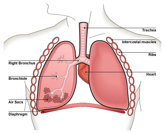
Lungs are the major organ of our respiratory system. They help you obtain oxygen and get rid of carbon dioxide. The gaseous exchange takes place in the tiny air sacs which have just one-cell thick walls. Blood absorbs oxygen and delivers it to every individual cell in the body.

A cylindrical tube, called trachea, is the main link between the human lungs and the nose and mouth. When we breathe in, air travels through the trachea to the main respiratory organs. As the trachea moves down the chest, it splits into two branches, i.e. bronchi, one for each lung.

Each bronchus then further subdivides into several smaller tubes, namely, bronchioles. Thus, each lung contains many bronchioles. They spread all over its structure in order to ensure an even distribution of oxygen. You can view the details in the lungs diagram.

For oxygen to be able to reach all the parts of our body, it dissolves into the blood. The blood stream then distributes oxygen to the entire body. Therefore, there has to be a supply of blood vessels in the lungs. These blood vessels are present on minute, sac-like structures, the alveoli.

The alveolar sacs are present at the end of bronchioles. There are numerous alveoli in each lung. This is to provide the maximum surface area for the exchange of oxygen as well as carbon dioxide with the blood stream.

Do you know the expanse combined surface area of all the tiny alveolar sacs in the lungs? Quite unbelievably, it is equal to the area of an entire tennis court.

It is possible for a person to survive with just one lung. However, he would be weaker than normal individuals who have a pair of these organs.

On average, the volume of air that the respiratory system takes in every day is approximately 9,000 liters!

The pulmonary structures of newly born babies breathe at a faster rate in comparison with those of adults.

Your pulmonary organs play an important role in respiration system of the body. Oxygen moves in when you inhale air. The air travels from the external air passages (mouth and nose) down the trachea and finally into the alveoli through the bronchus and bronchioles.

This oxygen dissolves into the layer of moisture around each alveoli and finally moves into blood capillaries. In this way, a lung plays a vital role in providing oxygen to all the body organs.

It is important for your body to get rid of carbon dioxide which is a waste product of respiration. Once again, these are the pulmonary organs which help in the elimination of this toxic waste product. The path of carbon dioxide from the blood to the external environment is the reverse of the path that the oxygen takes to enter the blood stream.

Sunday

Kidneys

Location of kidneys is of particular importance in your body. There are two bean shaped kidneys in the human body. A normal kidney is about 2.5 cm thick, 10 cm long and 5 cm wide. They are reddish-brown in color. Each weighs around 130 gm in adults. Both kidneys receive blood from the renal artery. In a resting adult nearly 1.2 to 1.4 liters of blood flows through both kidneys in one minute. It means every 4 to 5 minutes all the circulating blood in the body passes through the kidneys. Kidneys help remove excess salt, water and waste products from the bloodstream.

Like other endocrine organs, kidneys also secrete important hormones such as rennin and erythropoietin into the circulation. These hormones play a significant role in the production of blood cells. In addition, they also assist in the regulation of blood pressure.

It won’t be wrong to call kidney as the natural filter in your body. Its job is to purify blood and make it free from all the waste substances that it has collected from every individual cell. but how is such an important kidneys function executed? For this purpose, you need to study the kidney anatomy.

Location Of KidneysYou can better understand the anatomy of kidneys from the labelled diagrams. One of these is shown here. In the kidney images, you can see that a nephron serves as the basic structural and functional unit. There are a large number of nephrons in each of these organs. They make a direct connection with a complex network of blood vessels.

From the kidneys pictures, you can easily distinguish among different parts, such as medulla, cortex and the renal pelvis. On the side of the renal hilum, you will come across renal artery and the renal vein. From the renal pelvis, there emerges a ureter which descends downward to reach bladder. It also indicates that the location of kidneys is directly above the bladder.

The function of kidneys is to produce urine by filtering large quantities of fluid from the blood plasma. Urine consists of approximately 95% water. Urine also contains urea, creatinine, phenols, phosphate, potassium and sulfates. There are also nitrates and a large amount of uric acid.
Urea, uric acid and creatinine are all nitrogenous waste products. Nitrogen containing compounds are among the most toxic metabolic waste substances. Your body produces them as a result of various metabolic processes.

You can express the level of nitrogenous waste products in our blood as BUN (blood urea nitrogen). In healthy adults, normal BUN level ranges from 8 mg/dL to 25 mg/dL. A high level of BUN indicates renal insufficiency, i.e. one of the kidney problems.

The volume and composition of urine that the kidneys produce and excrete every day depend crucially on various factors. These factors include fluid intake, diet, renal functions, climate, metabolic activities of cells. Meanwhile, the presence or absence of systemic diseases also has a role to play in this regard.

Thursday

Gall Bladder


What is gallbladder? The gallbladder facts reveal that it has to play an important role in the breakdown of food items and, consequently, their absorption into the blood. Before learning gallbladder definition, you should know about the extent of digestive system. The digestive system is perhaps the largest system in the human body. It has many accessory organs and glands to perform its overall digestive function
According to gallbladder definition, it is a small gland at the base of liver. Its main job is to assist in the emulsification, digestion and absorption of fats in food. The liver secretes the bile and transfers it to gall bladder for temporary storage.

Another gallbladder function is to concentrate the alkaline fluid that contains salts, pigments, fats and about 85% watery component.

Recent gallbladder information suggests that this structure in an adult human would measure about 8 x 4 cm or a bid more. It secretes bile juice to facilitate digestive processes of oily diet. The organ has divisions into three parts, which are neck, body and fundus. The neck of gall bladder establishes a connection with liver through common bile duct and transcends its secretions through it. Fundus appears at the opposite end of this pear-shaped organ, while body lies in its proximity away from the neck.
Bile Juice Secretion & Storage: The liver secretes the bile juice and pours it into gallbladder. The gallbladder then stores the bile juice for some time.

As the need arises, the bile juice enters into the duodenum of small intestine. This commonly happens after taking food that is rich in cholesterol or other lipids.

Breakdown and Digestion: Do you know the bile from the gall bladder emulsifies fat molecules? As a result, the fat digesting enzymes, like lipase, can carry out their breakdown and digestion effectively.

Bile Concentration: Here is another of the interesting facts about the gallbladder in the digestive system. That is, the biliary vesicle achieves about 10 times concentration of bile.

Formation of Gallstones: Frequent and prolonged intake of fatty foods, would result in excessive concentration and hardening of bile juice. It will, in turn, result in the formation of gallbladder stones.

Gallbladder Removal: Removing gallbladder does not interfere much with fat metabolism and there direct secretion of bile into the intestine.

Inflammatory disorder: Inflammatory disorder of the gallbladder is also common among people who have been suffering from chronic infection of typhoid.

The gallstone are usually small, hardened deposits that measure not more than a few millimeters. That is why, in most of the cases, they do not cause any severe symptoms and go without detection for years. At the later stage, however, they lead to pain, infection and inflammation of the organ.

The gallstones can be of two major types, i.e. pigment stones and cholesterol stones. The pigment stones result from the accumulation of calcium bilirubinate – a chemical produced after the breakdown of red blood cells. In the latter case, accumulation of cholesterol is the sole cause for the formation of such stones.

There are usually very rare cases of this type of cancer. However, there are chances for its spreading to other parts of the body. In that case, the disease becomes fatal. If there is timely discovery of the gallbladder cancer, it is easily treatable. But, unfortunately, it is one of the gallbladder facts that in most of the cases, the cancer is diagnosed at a very later stage. The primary reason behind delay in the diagnosis is that such type of cancer does not cause any obvious signs or symptoms soon after its onset.

Saturday

Bronchi

Do you have any brief idea about “What is bronchi?”. Well, it is a plural form of bronchus. Here it becomes pertinent to define bronchus. A bronchus, on the other hand, can be defined as either of the two branches of trachea which lead directly to the lungs. So, the appendix definition makes it clear that a bronchus is actually a sub-division of the windpipe. The trachea or windpipe serves as a passage for airflow between mouth or larynx and the lungs.

Structurally composed of cartilaginous material, the air passages show a significant amount of flexibility. This flexibility, in turn, substantially contributes to their functioning efficiency. Any abnormal condition in the structure gives rise to great disturbance and severe symptoms of the respiratory system. But a number of remedial strategies are available for almost all of such disorders.
                          
Though their walls do not let the respiratory gases pass across them, the bronchial canals are the main channels for the passage of airstream towards the major respiratory organs. They also make significant contribution to the protection of lungs against germs and dirt particles. The germs and dirt particles are trapped in the mucous lining of the lumen.

Afterwards, there is upward expelling and removal of such unwanted things out of the windpipe with the help of ciliated cells. The hyaline cartilaginous material that forms the D-shaped incomplete rings of the bronchial walls prevents the tubes from collapsing and obstructing the flow of air.

However, with the passage of time as you grow older, this soft flexible material becomes a hard and rigid bony substance. Consequently, it reduces the overall efficiency of the structure. The alveolar sacs (or alveoli) form as a result of many subdivisions of the mainstem bronchi. And these are the places where the exchange of respiratory gases takes place.

There are a number of respiratory disorders that affect the subdivisions of windpipe at different levels, i.e. bronchi, bronchioles and alveoli or respiratory sacs. For example, bronchitis – as the very name suggests – is the inflammatory disorder of bronchi. It may occur in either of the two major forms, viz. chronic and acute one.

Chronic bronchitis results from the prolonged exposure to the irritants and smoking habits. While acute form is the result of the bacterial and viral infections. As a response to the allergens (antigens producing abnormally vigorous immune response), hyperactivity occurs in the bronchial canals. People call this condition as asthma.

Because of its vertical position, the right main bronchus is more vulnerable to the disorders. It is because the germs and dust particles present in the atmospheric air get entry into the right lung quite easily. Over there, they cause mild or severe medical conditions.

Wednesday

Appendix

Before turning to interesting appendix facts, let’s have basic information about this vestigial organ of the body. What is appendix definition? Literally speaking, an appendix is something added on to something else as an appendage. Are you really confused? Have you seen the appendix in the books? The appendix of a book is a collection of specific pages, containing additional information, in the end of the book. In this case, the appendix is added to the regular pages.

According to biological appendix definition, it is a finger like growth at the junction between small and large intestine. It has a blind end which means anything entering into it cannot come back. This organ is of no particular use to humans. However, appendix is still functional in the grazing animals.

It is located at the junction where small and large intestines join each other, i.e. in the right lower abdominal quadrant. Find it somewhere between the top section of the pelvic bone and the navel area. Normally, we cannot determine the exact location of the appendix and even in the scanned image.

However, it is not clearly visible. You can locate this body organ in the specified abdominal chamber,
when the visceral organs in human body are at their normal positions. Physicians refer to such a condition as situs solitus. There is a congenital condition (i.e. situs oppositus) in which there is delocalization or reversing of the internal organs. So, in the rare cases of situs inversus, this vestigial
organ appears on the opposite side of the body.

In human body, the vestigial structure of appendix develops a collection with the cecum of colon. Do you know what side is your appendix on? You can find this tube-shaped pouch in lower right side of your abdomen. It is very small in size and looks like a worm. That is why, you may also call it "Vermiform Appendix" or simply "Vermix" which comes from Latin meaning "worm-shaped"

Resembling a small, hollow, finger-like tube, vermix is blind at one end while the other end opens into cecum of large bowel (or colon). On average, the length is about 11 centimeters, but it varies greatly from person to person, and can enlarge up to 20 cm.

As the free end has no opening, anything entering into it will remain there with no way to come out of it. The stagnation of food will cause inflammation of the organ along with severe pain. Physicians can relieve the symptoms only with its immediate surgical removal, or else the victim may die. 

A well-known disease of appendix is appendicitis. Do you know the appendix causes? In this case, some food particles, passing through gastrointestinal canal, get an accidental entry into this hollow structure with blind end. But there is no way for the food to come out of it.

Thus, the stagnation of organic material produces pus and toxic substances. Consequently, it leads to severe inflammation. If you do not address the condition timely, the swollen tube bursts open. It releases its harmful contents to the sensitive internal organs, and may even cause the death of the victim.

Tongue

Did you ever think that, without the tongue, you would have been unable to taste all the deliciousness of the natural world? Can the scientists create and install such a tool in your body which would make you taste things in the absence of tongue? Maybe the invention of such an instrument is possible, it can never work as perfectly as your natural human tongue.Taking into account the value of human tongue, this small muscular organ in your mouth cavity is a wonderful blessing of nature. In addition to providing a taste of the food, the tongue helps in manipulation of food for mastication. Its third important function is to help in the swallowing of meal.
 As you can observe in the tongue pictures, the human tongue has got a rough texture. It is the presence of tiny bumps, called papillae, all over the dorsal surface of the tongue that make it look so rough. The surfaces of the papillae, on the other hand, contain thousands of taste buds. The taste buds are actually a collection of the nerve-like cells. Nerves develop a connection between taste buds and the brain.

The upper most layer of tissue on the human tongue is that of mucosa. The mucosa keeps it moist. It also makes the tongue a bit slippery to touch.

The ability of tongue to taste food is because of the presence of taste buds on its surface. Meanwhile, different parts of the tongue sense only a specific taste. Different types of receptor cells help to distinguish between various sorts of differently tasting substances, such as sweet, sour, bitter, spicy and salty. On the tip, you will find taste buds for sensing sweet. Bind the apex and along the sides, there are receptors for tasting bitter, sour and salt.

Dorsum – the upper surface of the tongue – contains lots of lingual papillae. The papillae house the taste buds and the taste receptors. These receptors assist in the sensation of taste of different types of dietary components which come into contact with them. They are basically of two types, that is, sweet taste receptors and bitter taste receptors.

In addition to enabling you sense the deliciousness of the food, the human tongue also performs a number of other important functions. For example, the additional tongue function involves the mastication of food and the act of swallowing it. Do you know any other interesting tongue function? The list of the some of the additional functionalities of the tongue is given below:

Thursday

Nose Definition

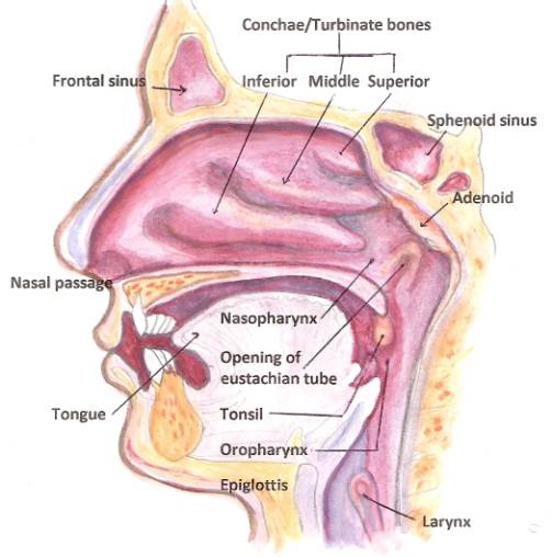
There are lots of amazing human nose facts which you may not be aware of. Also the human nose has got incredible importance in the life of an individual. For example, according to the nose definition, it serves as a passage for the entry of fresh air into your body. In other words, function of nasal passage is to assist in the process of respiration.
Human life will cease to exist in earthly atmosphere without fresh air. You get oxygen from the fresh air through nose. Here, you can call oxygen as the elixir of life. Across civilizations, man has been in search of good air.

In other words, healthy air is a primitive physiological need for humans. They base their existence on it on earth. But they failed to realize the importance of nose in human body. In addition to useful information, you will also come across various nose facts in this article.

The function of nose in respiratory system is very important. It is responsible for choosing, filtering and inhaling the atmospheric oxygen. The incoming oxygen is in sufficient amount to meet body needs. This article also throws some light on the basic parts of the nose and their functions.

Contributing to Facial Beauty:
Regarding it function, here are some interesting nose facts. Nose is the most prominent and perhaps the first attractive feature of the face. It occupies the central position along the geometrical division of the face. The organ has two chambers which divide in proportion by a septum.

These are the two nose parts. The human nose structure and function is of special interest for the biology students. It truly contributes to the overall beauty of humans.

After its purification, the air enters the middle nose for humidification. Meanwhile, the nasal cavity also warms the air to match body's internal temperature. Here is one of the wonderful nose facts. Before you accuse someone for not trimming his nasal hairs, remember they are inhaling better air than the rest of us.

⇛ A nasal septum separates the nasal cavity to form two equally dividing nostrils.

⇛ The fine hair or cilia in the nose collectively behave like a mechanical filter.

⇛ The atmospheric air is pre-treated to match body's temperature during inhalation.

⇛ The nose and the eyes are made of ethmoid bone. The ethmoid bone is present now
here else in the body other than the eye orbits.

⇛ One of the little interesting nose facts is the presence of Little's area at its interior aspect. This area may cause serious bleeding of no specific reason with constant picking of the nose.

⇛ Depending on their race, the individuals may lose their sense of small with age.

⇛ Which is the oldest sense in your body? Well, it is smell!

⇛ It may be one of the strange nose facts that women have a better sense of smell than that of men.

⇛ If someone has a long nose, they will show a good sense of business and respond positively to your leadership.