Tuesday

Lymph Nodes


They behave like mechanical filters. The job of these mechanical filters is to kill and eliminate microbes present in the blood stream. The white blood cells (WBCs) or lymphocytes are produced by the lymph nodes. On the other hand, WBCs kill bacteria and viruses entering the body.

Lymph nodes are very small organs, measuring only about 1-2 cm in size. They form a network throughout the body. There are about 500 lymph nodes spread across various organs of the human body.

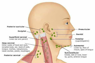
A lymph node gets its particular name from its location and relation with the body organs. For example, the cervical lymph nodes are present in the neck or cervical region of the body. They protect the tonsils and the pharynx. Here you should note that swollen cervical lymph nodes is an indication of the lymphatic system disease.

Lymph nodes in armpit are the axillary lymph nodes. Their job is to drain the chest region. You will find the mediastinal nodes near the sternum and the lung sacs. Meanwhile, for the drainage of the abdominal organs, there are the mesentery nodes.

Lymphatic System a Sewerage System: It won’t be wrong to compare the lymphatic system to a sewerage system. It has a networks of pipes to drain waste from every single body cell. The pipes in this system are defined as lymph vessels. The job of these vessels is to transport clear interstitial body fluid, that is, the lymph.

The Function of Lymph: The lymph is a watery fluid that flows in the lymphatic vessels. Its function is to help in effective draining mechanism.

Coordination of Drainage Mechanism: It is the muscles movements that coordinate the entire mechanism of draining. It is particularly because the lymphatic system has no specific pumping organs.

Bulging of Lymph Nodes: The lymph nodes bulge out in the presence of an infection. It is because, in case of an infection, they have to work hard to get rid of it.

Complementary Organs of Lymph Nodes: There are many organs in your body which protect the lymph nodes. They include spleen, tonsils, thymus and the adenoids. In addition to playing a protective role, these orangs are also complementary to the lymph nodes to form the immune system of body.

Lymphatic Circulation: About 2 liters of lymph fluid circulate in the body every day. During circulation, the lymph collects, destroys and eliminates foreign agents. Meantime, it also deals with the disease causing microbes.

Types of Lymphocytes: There are two types of lymphocytes or white blood cells that the lymph nodes produce. These are the B & T lymphocytes. The B-lymphocytes target bacteria and some viruses. Whereas the T-cells attack viruses in your body.
Monday

Parathyroid Gland

A number of vital functions in human body are controlled by biochemical substances, called hormones, which are chemical messengers released by either ductless or ducted glands directly into the blood stream or through ducts. Of all the hormone secreting glands in the body, the parathyroid gland is the smallest which is embedded in the thyroid gland or located on the rear side of it in the neck. There are four parathyroid glands located on the posterior side of the thyroid, measuring only about the size of a rice grain.
Because of the very small size, these endocrine glands weigh as low as 20mg or as high as 40mg. Usually, these are four in number, arranged in two pairs-one above and the other below-but some individuals have also been reported to have 6, 8 or even more. The two glands lying towards the head are termed as the superior parathyroid glands, while the other two lying on the lower side are known as the inferior parathyroid glands. Though the parathyroid glands are named after their close proximity with the thyroid gland, their functions are completely different from that of the neighbouring endocrine organ. Nervous and muscular systems are very sensitive to the concentration of calcium in the body that has to be maintained in a narrow range for their proper functioning. The hormone secreted by parathyroid gland (named after the gland as 'parathyroid hormone' or simply "parathormone") is responsible for the maintenance of phosphate and calcium homeostasis in the body, and is functionally opposed to calcitonin.

Though there are no pills or medications to cure the diseases of parathyroid gland, surgery and some other treatment measures have shown promising results.

Most of the parathyroid problems present themselves in their advanced stages, and some patients will not complain of any symptoms, until they are identified during routine diagnostic procedures.

Often the medical fraternity get confused with the diseases of the parathyroid gland and the blood calcium levels, as neither of these the conditions (diseases and calcium levels) have ever been found to synchronize with each other.

Most of the patients suffering from parathyroid disease have fluctuating blood calcium levels both in blood and bony structures.

The main function of the Parathyroid gland is to control the blood calcium levels within the range of 9-10 mg/dl which is done by the gastrointestinal absorption of calcium and its conservation in kidneys. It stimulates absorption of calcium from food and restricts kidneys from excreting excessive calcium in urine. If the blood calcium levels are low, the hormone would erode the bones to manage optimal blood calcium levels, thus making the bones weak and easily breakable. The gland also checks and regulates the levels of calcium deposition on the bones and their density.
Calcium not just makes the density of the bones and frames the skeletal system of the body, but is also vital to regulate and mediate nerve conduction and control of muscular activities (muscle contraction and relaxation). The electrical signals travelling from the tissues towards the master organ of the body (brain) are mediated with the help of calcium ions. The people who have blood calcium levels below the normal limits experience a sensation of prickling or extra-chill in their fingers along with severe muscular cramps in their legs.

Thursday

Salivary Glands

Concerning the salivary glands definition, these are glandular structures present at various locations in and around the oral cavity, they are responsible for the secretion of important digestive enzymes and fluids that help in the digestion and lubrication of dietary substances in your mouth. Ptyalin or salivary amylase and lipase are secreted by these ducted exocrine glands and are responsible for the oral digestion of food. Composed of cluster of secretary cells, called acini, they also synthesize mucous, water and electrolytes that are then released into the collecting ducts. Among the different types of salivary glands, submandibular, sublingual and parotid are more important as each of which is assigned a particular task to perform. The large particles of food are ground into smaller ones that are then moistened and converted into a semi-solid mass of partly digested food (almost round in shape) which you tem as 'bolus'. Some painful medical condition may arise out of the malfunctioning of salivary glands which cause severe disturbances in the region but can easily be relieved through various treatment measures.

Based on their function and particular location in and around the mouth cavity, the saliva secreting organs can be divided into different types, viz. sublingual glands, submandibular glands, Von Ebnor's glands, parotid glands and minor salivary glands. Parotid is present behind the mandibular ramus and constitutes the largest of salivary glands that releases its serous secretions into the oral chamber through Stensen's duct. Pouring the mucus and serous fluids into the mouth through Wharton's duct, the submandibular glands are found beneath the jaws. As the very name suggests, sublingual glands are located beneath the tongue and contribute about five percent of the salivary secretions in the oral chamber. Scattered across mucosa in the mouth, there are about 800 to 1000 minor salivary gland parts or that are surrounded by connective tissue and secrete mucous that is delivered to the oral cavity through a shared excretory duct.

Friday

Spinal Cord


Measuring up to 18 inch in length, the dorsal neural tube gradually decreases in width while running downwards in the vertebral column, i.e. at the cervical regions the thickness is about 1/2 inch while reaching the lumbar vertebrae the size is reduced to just 1/4 inches in diameter. However, it is comparatively shorter in length than your bony spinal column and does not run along its entire length. On the whole, the ovoid structure of neural tube can be subdivided into 33 segments in human beings. Based on their association with the different regions of vertebrae, these segments can be grouped into five broader categories which are briefly described as under:

Cervical Segments

Being the anterior most segments of the spinal cord that run across seven cervical vertebrae, it is located along the neck region. The upper portion forms as many as eight pairs of cervical nerves which are named with the vertebra they emerge from, viz. C1, C2, C3, C4, ? C8. However, the C8 pair of cervical nerve emerges from the lower end of the C7 vertebral segment while all of other pairs emerge from the upper end of the respective vertebra.

Thoracic Segments

Running along the series of twelve T vertebrae in the thoracic region, they constitute 12 pairs of thoracic nerves each of which leaving the vertebral column below the associated vertebral division.

Lumbar Segments

It is one of the parts of spinal cord that is protected by five lumbar vertebrae, it gives rise to five pairs of nervous extensions each of which emerges below the associated vertebra.

Control of Reflexes
Another major task of this part of your CNS is the independent control and coordination of central nerve generators and certain reflexes. Such actions protect your body against dangerous stimuli by the generation and application of a quick response. Under such circumstances, the prolongation of response may inflict an irreparable damage to your body.

Ailments of the spinal nerve column are generally referred to as the disorders of central nervous system as it is its second major component. Besides injuries, a number of diseases have been identified affecting the region, viz. infections, inflammations, tumors, and degenerative disorders. Such spinal abnormalities are revealed through different clinical manifestations, like the loss of sensation, numbness, severe pain, and muscle weakness. The symptoms usually occur around the spinal cord but may also spread to arms and legs. For their amelioration, your health care provider may advise you different treatment measures mainly depending on the type and severity of the disorder, but the most frequently recommended strategies include drug administration and surgical therapy.

Tuesday

Trachea

Concerning trachea definition, it is an important anatomical structure that is an integral part of respiratory system in humans and other higher animals. Allowing the passage of air (respiratory gases) it connects larynx (voice box) and pharynx with lungs. The trachea location can be traced in the chest region, and it is made of soft bony material, called cartilage that allows great amount of flexibility for the efficient performance of its essential tasks. However, as the age progresses, it becomes rigid and hard, and is converted into bone. Apart from just gaseous exchange, it is also involved in the production of vocal sounds as one of the secondary trachea functions. As with other organs of your body, trachea is also vulnerable to a number of diseases that are commonly known as respiratory disorders, viz. tracheal stenosis, tracheobronchomalacia,

As you can see in the trachea pictures, the windpipe is structurally composed of incomplete rings, resembling the letter C in English which range from 15 to 20 in number. At the anterior end, the air passage is bound by a membranous structure, called larynx that contains vocal chords running horizontally across its mouth and forming voice apparatus in humans for the production of speech sounds. At the posterior end, it undergoes dichotomous branching, thus dividing into the two smaller tubes, viz. primary bronchi. Each of the primary bronchi runs into either of the lungs (i.e. left or right one) and allows the smooth and efficient passage of air (or the exchange of respiratory gases). Going deeper into lungs, further branching takes place in which bronchi are subdivided into bronchioles ultimately attaining the size of very small air sacs enclosed by respiratory membranes through which exchange of respiratory gases (carbon dioxide & oxygen) virtually takes place. With the help of incomplete rings, the anterior of the tube (lumen) can be narrowed down to accelerate the air passing through it when needed, e.g. while coughing.

Any ailment of the respiratory system may disturb the overall functioning of your body as it is responsible for the facilitation of the process of energy extraction from food. Forming front tubular part of the respiratory system, the anatomical structure, if attached by some acute or chronic disorder, results in the partial or complete collapse of airway system and often leads to the death of the individual. It is surprising to know that this is the organ system of your body which is exposed and extremely vulnerable to environmental hazards, especially in this highly polluted atmosphere. A number of tracheal ailments may arise out of various factors, like infectious and hereditary which include inflammation, tumor formation, cancerous outgrowth, obstructive disorders, etc. Tracheal stenosis is characterized by inflammation of the windpipe that may be relieved by endoscopy or surgical therapy. Formation of large tumor (benign or cancerous) in the lumen of trachea may cause the blockage of the air passage and death of the victim due to chocking. Though it is very difficult to avoid contact with respiratory diseases, proper care, preventive measures and early treatment of the abnormalities can save you from life long miseries.

Sunday

Ears

As you can see in the human ear diagram, it is designed in such a way that it captures the maximum auditory stimuli from the atmosphere and transfers them to the brain for translating into hearing response. Three major parts of a human ear are the outer, middle and inner respectively, each having its own functions, anatomy and ear diseases as well.
The external ear anatomy is the study of the parts of the outer ear. There are two major parts of the external ear, i.e. the pinna and the external acoustic meatus. Pinna or the auricle lies externally and its main job is to capture the sound waves and transmit them to the external acoustic meatus. Most of the framework of the auricle is cartilaginous in nature. Here lobule is the only structure which does not have the support of cartilage.

The external acoustic meatus, on the other hand, is a tube like structure that extends from the middle hollow depression or concha of the auricle. At the other end, it reaches the tympanic membrane. It has a cartilaginous structure and gets the bony support from the temporal bone. The branches of vagus and mandibular nerves provide sensory innervation to this particular part of your external ear.

Do you know if the external acoustic meatus travel in the straight path? Well, this is not the case. In fact, it has got an S-shaped path.

⇛ Link between Cold and Dumbness: Do you know about one of the amazing ear facts that there is a connection between the middle ear and the throat? This connection develops through a tube, i.e. the Eustachian tube. This is the very reason behind your going dumb every time you catch cold. The main function of the tube is to equalize the pressure between the atmosphere and the body.

⇛ Disturbance in Equilibrium: At higher pressures, and on mountain tops, it is common for people to go dumb. Meanwhile, they experience a disturbing feeling in their ears. It is because the Eustachian tube is unable to maintain equilibrium in body pressure.

⇛ Strength of Temporal Bone: According to the opinion of many people, femur is the hardest, longest and strongest bone in human body. But it does mean that the temporal bone is less in strength, i.e. it is equally hard and strong.

⇛ Remove Wax & Damage The Ear: Ear produces its own wax to protect itself from friction and dust. So, if you make frequent attempt to remove this wax, it may damage the tympanic membrane in the ears causing deafness.

Do you know exactly how the ear works? The sound waves that strike at the ear drum set it on vibration. This vibration then travels to the three small bones of the middle ear, (the malleus, stapes and incus). The bones of the middle ear, on the other hand, make specific movements and hit at the oval window.
Different parts of the ear and their functions are very important for hearing. The vestibular window separates the middle ear from the inner ear. The inner ear is also termed as cochlea, which is a dual membrane delicate structure, with fluids filled between the membranes and lined with hair cells. The movement of the hair cells that line cochlea, in response to the sound waves, is like that of grasses in the meadows. It serves to sends electrical impulses to the brain, i.e. the master organ, that accurately identifies the sounds we hear.

Tuesday

Rectum

The straight part of the colon is the dilated pouch like structure that is preceded by sigmoid colon and followed by anal canal. Before you defecate, the feces is temporarily stored in this gastrointestinal structure which becomes dilated on the arrival of waste material and induces the urge for its elimination through anal verge. The word "rectum" has been derived from a Latin phrase "Rectum Intestinum" which means "straight intestine". As the rectum facts suggest, this organ is about 12 cm in length, and begins at the third sacral vertebrae and is joined with the preceding section of large bowel at rectosigmoid junction. Based on the structure and function of the distal end of large gut, you can divide rectum parts

The primary rectum function is the temporary storage of stool so that you may have some time to reach the place where it can be defecated. As the waste and indigestible materials get entry into it, the canal becomes dilated, thus stretching the muscles lining the rectal region. Stretch receptors are located in the wall of rectum which sense the dilation of the gut and send signal to the nervous system (brain) where these are processed and a response is generated that induces the need for the elimination of waste materials passing through anal canal and getting out through anal verge. However, if you do not go for defecation for a prolonged duration, the stool gets back into the colon for further absorption of fluids which may also result in hardening of stool and constipation.